Por favor, use este identificador para citar o enlazar a este item:
http://hdl.handle.net/10261/265823COMPARTIR / EXPORTAR:
|
|
|
| Visualizar otros formatos: MARC | Dublin Core | RDF | ORE | MODS | METS | DIDL | DATACITE | |

| Título: | Metallated nanoparticles as dual-imaging theranostic system in a glioblastoma mouse model |
Autor: | Serrano-Torres, María CSIC; Arias-Ramos, Nuria CSIC ORCID; Ibarra, Luis E.; Guillén Gómez, María José CSIC; Caverzán, Matías Daniel; Palacios, Rodrigo E.; Chesta, Carlos A.; López-Larrubia, Pilar CSIC ORCID | Fecha de publicación: | 7-oct-2021 | Citación: | ESMRMB 38th Annual Scientific Meeting (2021) | Resumen: | [Introduction]: Glioblastoma multiforme (GBM) is considered the most lethal of the malignant primary brain tumors [1]. In fact, even after aggressive treatment, survival rates are approximately 12-15 months after diagnosis [2]. Recently, nanosystems have become promising candidates for GBM diagnosis and treatment, due to their exceptional magnetic properties, biocompatibility and blood brain barrier (BBB) penetrability [3]. In our previous investigations, metallated doped conjugated polymer nanoparticles (CPNs) (conjugated with fluorescent polymer F8BT) were visualized in tumors by T2 weighted (T2W) MRI [4]. In this sense, this project aims to evaluate the biodistribution of two CPNs with different types of cores, Fe3O4 or NiFe2O4, in mice bearing GBM flank-tumors and control mice. [Methods]: In vitro validation of both CPNs was performed in a phantom study with different CPNs dilutions (Fig. 1A). Afterwards, NOD-SCID mice were injected intravenously with CPNs with a Fe3O4 or NiFe2O4 core. CPN’s biodistribution was studied by T2W magnetic resonance imaging (MRI) pharmacodynamics (Fig. 1C & D) and T2 maps (Fig. 1B) were obtained before and after the CPNs injection in a 7T system. An hour after injection, mice were sacrificed, organs were resected and studied by fluorescent imaging. Additionally, mice bearing C6-GBM flank tumors were studied by T2W MRI before and 15 minutes after intertumoral injection Fe3O4 or NiFe2O4CPNs. Then, mice were sacrificed and flanks removed for fluorescence studies using a IVIS Lumina II system. [Results]: We observed a higher CPNs uptake in the liver and a moderate accumulation in the renal cortex and the renal medulla as seen in T2W pharmacodynamics (Fig. 1C & D). The CNPs liver accumulation seem to be higher with Fe3O4 than NiFe2O4 core nanoparticles (Fig. 1C). Intratumor injection studies revealed that both CPNs can be visualized in flank tumors by T2W images, showing signal decreasing in the location where the CPNs were injected (Fig. 2A). Both CPNs were also detected in the xenograft tumors by fluorescence imaging and not observed in not injected (control) tumors (Fig. 2B). [Discussion]: Overall, results suggest that both CPNs are good candidates to study whether they reach and accumulate in the tumor of an orthotopic and xenograft GBM model | Descripción: | Trabajo presentado en el ESMRMB 38th Annual Scientific Meeting, celebrado en modalidad virtual del 07 al 09 de octubre de 2021. | URI: | http://hdl.handle.net/10261/265823 |
| Aparece en las colecciones: | (IIBM) Comunicaciones congresos |
Ficheros en este ítem:
| Fichero | Descripción | Tamaño | Formato | |
|---|---|---|---|---|
| Metallated_nanoparticles_Serrano_Torres_Póster2021.pdf | 1,15 MB | Adobe PDF | 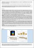 Visualizar/Abrir |
CORE Recommender

NOTA: Los ítems de Digital.CSIC están protegidos por copyright, con todos los derechos reservados, a menos que se indique lo contrario.