Por favor, use este identificador para citar o enlazar a este item:
http://hdl.handle.net/10261/92296COMPARTIR / EXPORTAR:
|
|
|
| Visualizar otros formatos: MARC | Dublin Core | RDF | ORE | MODS | METS | DIDL | DATACITE | |

| Título: | Placental thrombosis in acute phase abortions during experimental Toxoplasma gondii infection in sheep |
Autor: | Castaño, P.; Fuertes Franco, Miguel CSIC ORCID; Fernández Fernández, Miguel CSIC; Ferreras, Mª del Carmen CSIC ORCID ; González Lanza, Camino CSIC ORCID ; Pérez Pérez, Valentín CSIC ORCID ; Benavides, Julio CSIC ORCID | Palabras clave: | Acquired-immunodeficiency-syndrome Ovine toxoplasmosis Pregnant sheep Oocysts Vaccine Transmission Challenge Cells |
Fecha de publicación: | 29-ene-2014 | Editor: | BioMed Central | Citación: | Veterinary Research 45: 9 (2014) | Resumen: | After oral administration of ewes during mid gestation with 2000 freshly prepared sporulated oocysts of T. gondii isolate M4, abortions occurred between days 7 and 11 in 91.6% of pregnant and infected ewes. Afterwards, a further infection was carried out at late gestation in another group of sheep with 500 sporulated oocysts. Abortions happened again between days 9 and 11 post infection (pi) in 58.3% of the infected ewes. Classically, abortions in natural and experimental ovine toxoplasmosis usually occur one month after infection. Few experimental studies have reported the so-called acute phase abortions as early as 7 to 14 days after oral inoculation of oocysts, and pyrexia was proposed to be responsible for abortion, although the underline mechanism was not elucidated. In the present study, all placentas analysed from ewes suffering acute phase abortions showed infarcts and thrombosis in the caruncullar villi of the placentomes and ischemic lesions (periventricular leukomalacia) in the brain of some foetuses. The parasite was identified by PCR in samples from some placentomes of only one sheep, and no antigen was detected by immunohistochemical labelling. These findings suggest that the vascular lesions found in the placenta, and the consequent hypoxic damage to the foetus, could be associated to the occurrence of acute phase abortions. Although the pathogenesis of these lesions remains to be determined, the infectious dose or virulence of the isolate may play a role in their development. | Versión del editor: | http://dx.doi.org/10.1186/1297-9716-45-9 | URI: | http://hdl.handle.net/10261/92296 | DOI: | 10.1186/1297-9716-45-9 | ISSN: | 0928-4249 |
| Aparece en las colecciones: | (IGM) Artículos |
Ficheros en este ítem:
| Fichero | Descripción | Tamaño | Formato | |
|---|---|---|---|---|
| 1297-9716-45-9.xml | 82,88 kB | XML | Visualizar/Abrir | |
| 1297-9716-45-9.pdf | 1,72 MB | Adobe PDF | 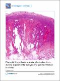 Visualizar/Abrir |
CORE Recommender
PubMed Central
Citations
19
checked on 09-abr-2024
SCOPUSTM
Citations
36
checked on 08-abr-2024
WEB OF SCIENCETM
Citations
31
checked on 28-feb-2024
Page view(s)
252
checked on 17-abr-2024
Download(s)
410
checked on 17-abr-2024

